Fig. 2.
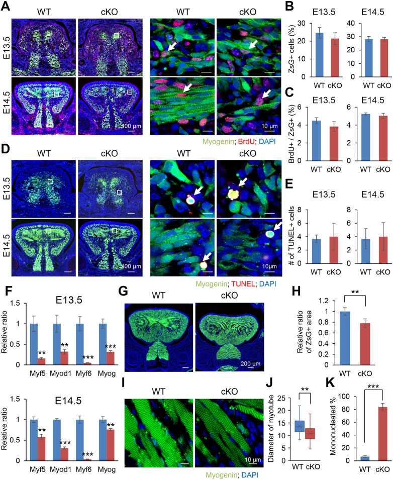
Loss of β-catenin in the Myog-Cre-positive lineage causes a defect in muscle differentiation. (A) BrdU staining of Myog-Cre;Ctnnb1F/F;ZsGreencKI/cKI (cKO) and Myog-Cre;Ctnnb1F/+;ZsGreencKI/cKI control (WT) embryos at E13.5 and E14.5. The boxed areas are enlarged in the right panels. Arrows indicate BrdU-positive cells (red). Myog-Cre+ myoblasts are green. Nuclei were counterstained with DAPI (blue). (B) Percentage of ZsGreen-positive cells in the tongue from Myog-Cre;Ctnnb1F/F;ZsGreencKI/cKI (cKO) and Myog-Cre;Ctnnb1F/+;ZsGreencKI/cKI control (WT) embryos at E13.5 and E14.5. (C) Percentage of BrdU-positive cells per ZsGreen-positive cells in the tongues from Myog-Cre;Ctnnb1F/F;ZsGreencKI/cKI (cKO) and Myog-Cre;Ctnnb1F/+;ZsGreencKI/cKI control (WT) embryos at E13.5 and E14.5. (D) TUNEL staining of Myog-Cre;Ctnnb1F/F;ZsGreencKI/cKI (cKO) and Myog-Cre;Ctnnb1F/+;ZsGreencKI/cKI control (WT) embryos at E13.5 and E14.5. The boxed areas are enlarged in the right panels. Arrows indicate TUNEL-positive cells (red). Myogenin-positive myoblasts are green. Nuclei were counterstained with DAPI (blue). (E) Number of TUNEL-positive cells in the tongues from Myog-Cre;Ctnnb1F/F;ZsGreencKI/cKI (cKO) and Myog-Cre;Ctnnb1F/+;ZsGreencKI/cKI control (WT) embryos at E13.5 (top) and E14.5 (bottom). (F) Quantitative RT-PCR analyses for muscle differentiation markers in the tongues from Myog-Cre;Ctnnb1F/F;ZsGreencKI/cKI (cKO) and Myog-Cre;Ctnnb1F/+;ZsGreencKI/cKI control (WT) embryos at E13.5 and E14.5. **P<0.01; ***P<0.001. (G) Fluorescent images from newborn Myog-Cre;Ctnnb1F/F;ZsGreencKI/cKI (cKO) and Myog-Cre;Ctnnb1F/+;ZsGreencKI/cKI control (WT) mice. Myogenin-positive myoblasts are green. Nuclei were counterstained with DAPI (blue). (H) Quantification of the ZsGreen-positive area in the tongue of Myog-Cre;Ctnnb1F/F;ZsGreencKI/cKI (cKO) and Myog-Cre;Ctnnb1F/+;ZsGreencKI/cKI control (WT) mice. **P<0.01. (I) Fluorescent images from newborn Myog-Cre;Ctnnb1F/F;ZsGreencKI/cKI (cKO) and Myog-Cre;Ctnnb1F/+;ZsGreencKI/cKI control (WT) mice. Myogenin-positive myoblasts are green. Nuclei were counterstained with DAPI (blue). (J) Diameter of ZsGreen-positive myotubes in the tongues of Myog-Cre;Ctnnb1F/F;ZsGreencKI/cKI (cKO) and Myog-Cre;Ctnnb1F/+; ZsGreencKI/cKI control (WT) mice. **P<0.01. (K) Percentage of mono-nucleated myotubes in the tongues of Myog-Cre;Ctnnb1F/F;ZsGreencKI/cKI (cKO) and Myog-Cre;Ctnnb1F/+;ZsGreencKI/cKI control (WT) mice. ***P<0.001. Scale bars: 100 µm in A,D (left); 10 µm in A,D (right),I; 200 µm in G.

