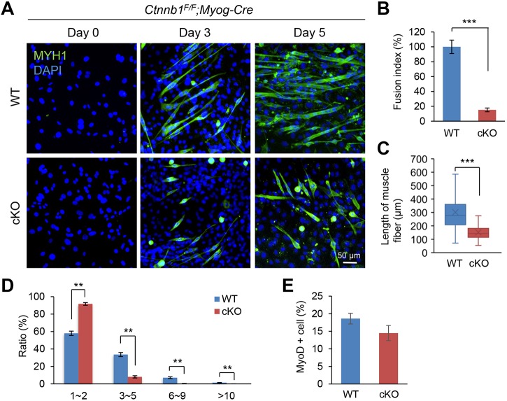
Fig. 3.
Ctnnb1F/F;Myog-Cre myoblasts show a fusion defect during muscle differentiation. (A) Myoblasts from wild-type (WT) control and conditional null (cKO) mice were cultured in muscle differentiation medium for the number of days indicated. Myotubes from WT and cKO tongues were stained with anti-MYH1 antibody (green), and the nuclei were stained with DAPI (blue). Scale bar: 50 µm. (B) Fusion index at day 5 of cultured cells from WT (blue bar) and cKO (red bar) tongues. ***P<0.001. (C) Length of muscle fibers in cultured cells from WT (blue bar) and cKO (red bar) tongues. ***P<0.001. (D) Ratio of muscle cells with the indicated number of nuclei in cultured cells from WT (blue bars) and cKO (red bars) tongues. (E) Percentage of MYOD1-positive myoblasts from WT control (blue bar) and cKO (red bar) tongues at day 0. **P<0.01.

