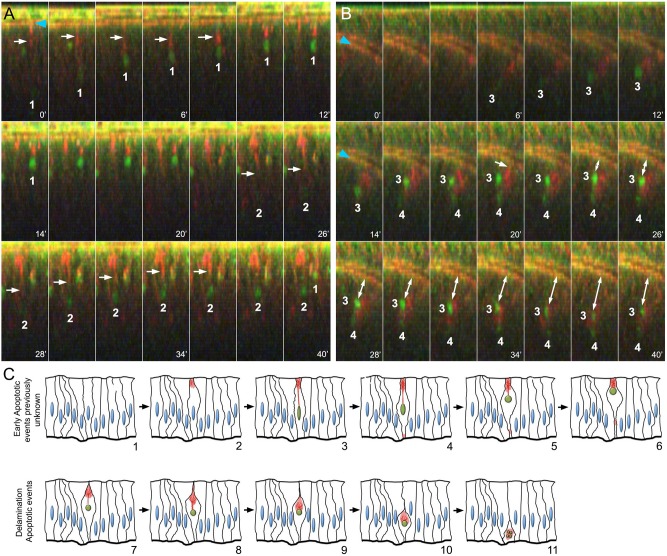
Fig. 2.
Live imaging of imaginal discs upon irradiation. (A) Time-lapse imaging of wing imaginal discs ex vivo (cross-sections) after irradiation (time is indicated in minutes; DBS-S is shown in green; lifeAct-Ruby shows actin in red). Notice the progressive accumulation of GFP signal in the nuclei of cells 1 and 2, the changes in their shape (roundness increase) and the movement towards the apical cell cortex. White arrows indicate the actin bundles connecting the apical cell cortex with the nuclei; notice the contraction of the actin bundle over time. The recording started 90 min after irradiation and lasted for 40 min. Snapshots were acquired every 2 min. (B) Delamination process of apoptotic cells 3 and 4 after completing apical migration; white arrow indicates apical cell detachment before delamination (20 min). Blue arrowheads indicate the normal accumulation of actin in the apical adherent cell junctions. Notice that actin remains accumulated around the nuclei during the movement towards the basal surface of the epithelium. Image acquisition conditions are described in A. Double-headed arrow indicates the basal delamination of DBS-S positive cells. (C) Schematic of early apoptotic events newly identified by DBS-S and lifeAct-Ruby in irradiated wing discs (1-6), as well as delamination events previously described during apoptosis (7-11). The activation of the reporter is indicated by the green nuclei, and the actin network is represented in red.

