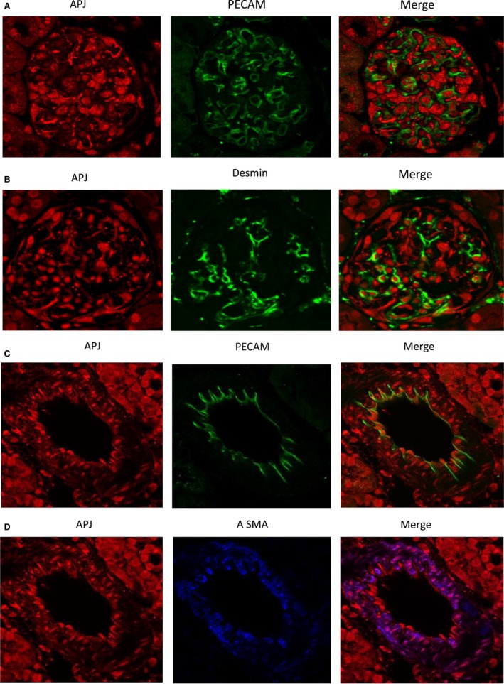
Figure 3.

Immunofluorescence staining of APJ (red; left) and the endothelial cell marker, the platelet‐endothelial cell adhesion molecule, PECAM‐1 (green; middle; A) and desmin (green, middle; B) in a kidney glomerulus. APJ shows no colocalization with PECAM‐1 (right, A) and little with desmin (yellow; right, B). Double immunofluorescence staining of APJ (red; left, C) and the endothelial cell marker platelet‐endothelial cell adhesion molecule PECAM‐1 (green; middle; C) and the vascular smooth muscle marker, the α‐smooth muscle actin, α‐SMA (blue; middle; D) in a renal vessel from mouse kidney. APJ shows no colocalization with PECAM‐1 (right), but colocalizes strongly with α‐SMA (pink; right).
