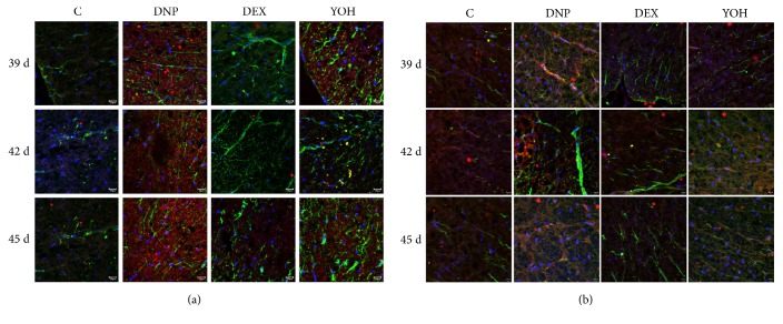
Figure 4.
Immunohistochemical analysis showing astrocyte activation and Wnt 10a and β -catenin localization. On days 39, 42, and 45, spinal cord sections were stained with anti-GFAP (green) antibody and anti-Wnt 10a (red) antibody (a). Nuclei were counterstained with DAPI. On days 35, 42, and 45, sections were stained with anti-GFAP (green) antibody and anti-β-catenin (red) antibody (b). n=4. C, control; DNP, diabetic neuropathy pain; DEX, dexmedetomidine; YOH, yohimbine.

