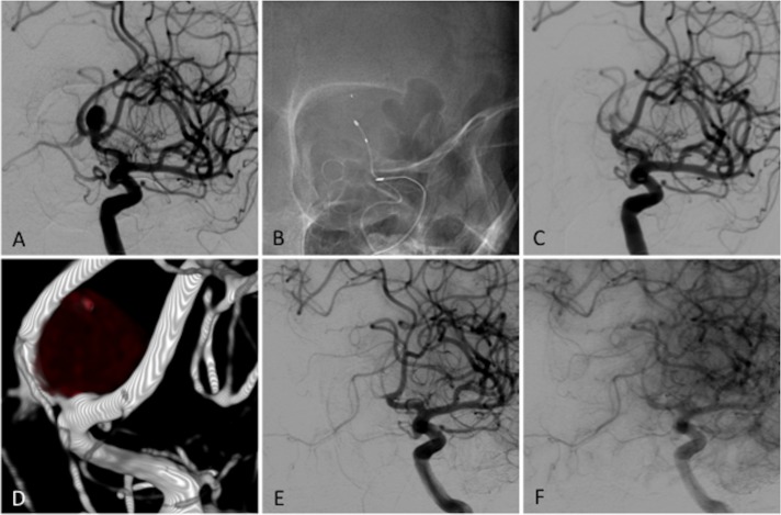
Figure 2.
Occlusion example. (A) Baseline angiography for a patient with a 8.0 mm anterior communicating saccular aneurysm. (B) Plain radiograph during the LUNA aneurysm embolization system (AES) implantation. (C) Immediate control angiogram after implantation showing flow reduction within the aneurysm. The same patient at 36-month follow-up with 3D angiography: (D) early arterial phase of angiography; (F) late arterial phase showing complete circulatory exclusion of the aneurysm.

