Figure 3.
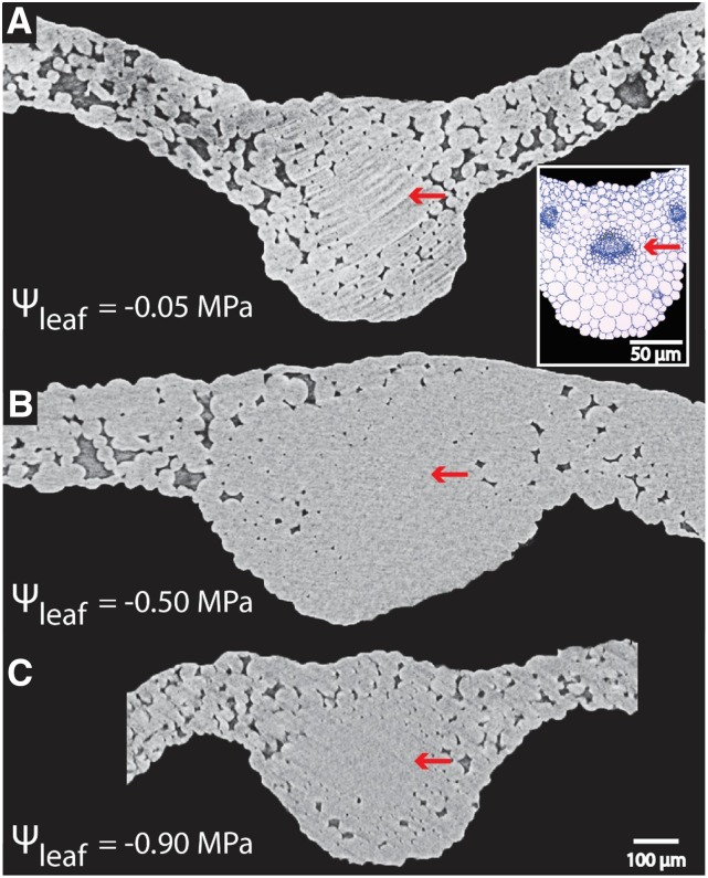
Lack of embolism observed in midrib conduits of Arabidopsis (Col-0) across levels of dehydration, as revealed by in vivo images of leaf midribs subjected to progressive dehydration using microCT. Water-filled cells appear in light gray in microCT. If air-filled (i.e. embolized) conduits were present, they would appear as black in the xylem portion of the midrib. There was no embolism, as shown in these images by the red arrows pointing at the entirely light gray midrib xylem. The Ψleaf has been provided for each image. The inset in A represents a leaf midrib cross section imaged under light microscopy, with the red arrow pointing to the xylem tissue (dark blue conduits).

