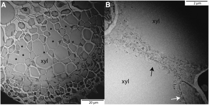
Figure 8.
Transmission electron microscopy of Arabidopsis (Col-0) midrib cross sections. In A, the entire xylem (xyl) portion of the midrib can be seen. Black arrows point to the lack of secondary lignified wall around xylem conduits. These long primary wall sections can be observed in more detail in B. The white arrow points to a lignified portion of the secondary xylem wall. We hypothesize that the xylem resistance through these deeply helicoidal xylem conduits is reduced greatly, as unlignified primary cells effectively work as one large pit membrane.

