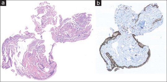
Figure 3.

Microbiopsy specimen ×20 original magnification. H&E stain (a) reveals fragments of mucinous epithelium with basally oriented nuclei. The epithelial cells are immunohistochemically positive for MUC1 (b), and MUC5AC indicative of intraductal papillary mucinous neoplasm
