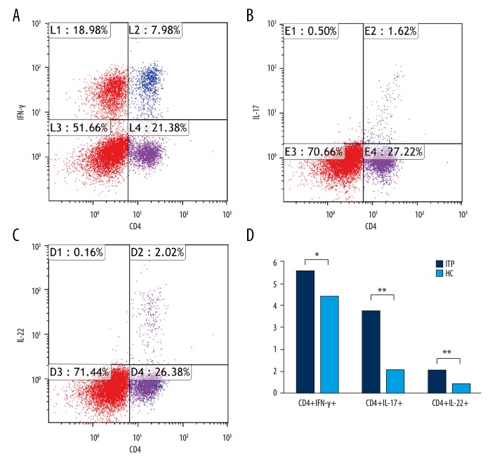
Figure 1.
Quantification of circulating CD4+IFN-γ+, CD4+IL-17A+, and IL-22+ cells in each group. (A–C) Representative four-color dot plot analyses of CD4+IFN-γ+, CD4+IL-17A+, and IL-22+ cells after stimulation with PMA and ionomycin. (D) Frequency of CD4+IFN-γ+, CD4+IL-17A+, and IL-22+ cells on the gated lymphocytes in the FSC/SSC plot in each group. Results are represented as the mean ±SD (** p<0.05). ANOVA followed by Tukey’s post hoc test was used to analyze statistical significance.

