. 2018 Nov 19;47(24):9069–9105. doi: 10.1039/c8cs00590g

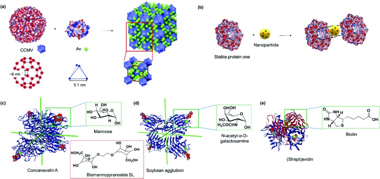
Fig. 2. (a) The electrostatic surface of native cowpea chlorotic mottle virus and avidin (Av) with the location and geometry of the surface patches, which allow interactions to form binary protein crystals. Adapted with permission from ref. 34. Copyright 2014 Nature Publishing Group. (b) The electrostatic surface of stable protein one (SP1, PDB: ; 1TR0) and the central cavity that interacts with polycationic nanoparticles with specific dimensions. Blue denotes positive surface charges, red denotes negative surface charges, and white refer to neutral areas. (c) Tetrahedral concanavalin A (PDB: ; 1CVN) versus (d) slightly out of plane soybean agglutinin (PDB: ; 1SBF) that assembles with sugar units. Red inset in (c) shows a bismannopyranoside SL. Both PBs in (c and d) were viewed along the D2 symmetry axes for direct comparison. (e) (Strept)avidin tetramer (PDB: ; 1MK5) with the binding ligand biotin. Insets in (c–e) shows the chemical structure of the biotin ligand. Protein images were created using the NGL viewer.240.

Fig. 2