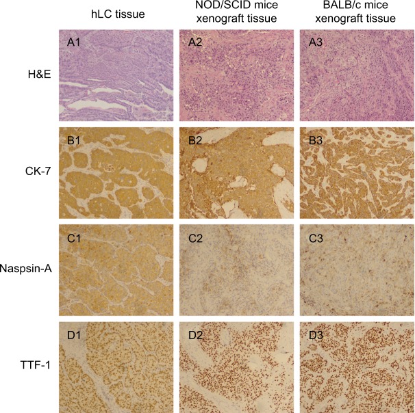
Figure 1.
Comparison of building PDX models in NOD/SCID mice and BABL/c mice with the corresponding adenocarcinoma patient.
Notes: (A1) H&E staining of human lung cancer tissue; (A2) H&E staining of NOD/SCID mice xenograft tissue; (A3) H&E staining of BALB/c mice xenograft tissue; (B1) CK-7 staining of human lung cancer tissue; (B2) CK-7 staining of NOD/SCID mice xenograft tissue; (B3) CK-7 staining of BALB/c mice xenograft tissue; (C1) Naspin A staining of human lung cancer tissue; (C2) Naspin A staining of NOD/SCID mice xenograft tissue; (C3) Naspin A staining of BALB/c mice xenograft tissue; (D1) TTF-1 staining of human lung cancer tissue; (D2) TTF-1 staining of NOD/SCID mice xenograft tissue; (D3) TTF-1 staining of BALB/c mice xenograft tissue. Magnification ×100.
Abbreviations: hLC, human lung cancer; PDX, patient-derived tumor xenografts.

