Figure 5.
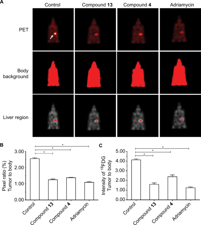
Compound 4 or 13 inhibited the MicroPET signaling mice with the intrahepatic growth of MDA-MB-231 cells in the liver region.
Notes: MDA-MD-231 cells were injected into nude mice’s liver via hepatic portal vein. Next, the mice were divided into four groups: 1) solvent control group; 2) compound 4 treatment group; 3) compound 13 treatment group; 4) adriamycin treatment group. The tumor nodules formed by MHCC97-H cells in mice’s liver were examined by MicroPET scanning. Results are shown as follows: (A) MicroPET results from animals, (B) pixel ratio of tumor to body, or (C) intensity of tumor to body. Arrow indicates the tumor tissues in the liver. *P<0.05.
Abbreviation: PET, positron emission tomography.

