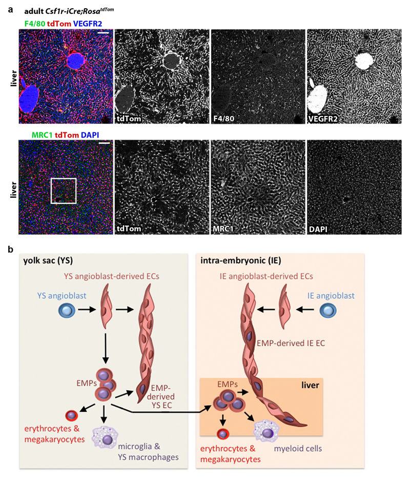
Extended data figure 10. Csf1r-iCre-targeted ECs contribute to adult organ vasculature.
(a) 20 µm cryosections of 3 months old adult Csf1r-iCre;RosatdTom livers (n = 3) were immunolabelled for RFP and the EC marker VEGFR2 and the macrophage marker F4/80 or the liver EC sinusoidal EC marker MRC1 and then counterstained with DAPI; single channels are shown in grey scale. The white box indicates an area shown in higher magnification in Fig. 6h. Scale bars: 100 µm.
(b) Working model for the role of EMPs in generating extra-embryonic yolk sac and intra-embryonic organ ECs alongside their known role in generating myeloid and erythrocyte/megakaryocyte cells.

