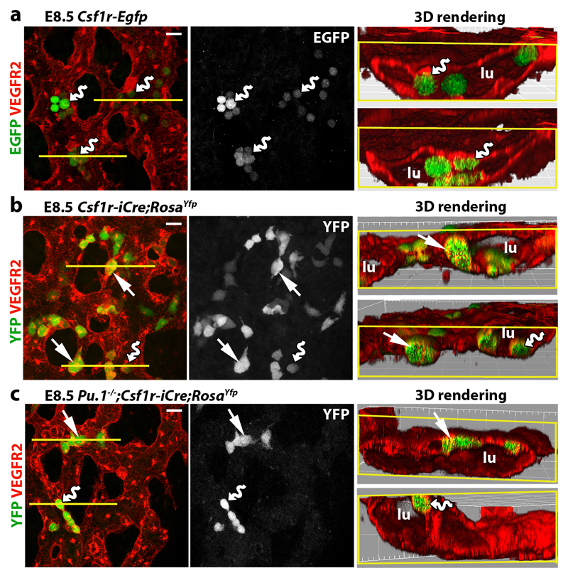
Fig. 2. Csf1r-iCre-targeted ECs emerge concomitantly with EMPs in the yolk sac.
E8.5 yolk sacs were wholemount labelled with the indicated markers. (a) Csf1r-Egfp yolk sacs. (b,c) Csf1r-iCre;RosaYfp yolk sacs on a Pu.1+/+ versus (b) Pu.1-/- background (c). N = 4 yolk sacs for each genotype. The yellow lines mark the start of 3D-rendered lateral views. Wavy arrows indicate VEGFR2+ EGFP+ and VEGFR2+ YFP+ round EMPs/MPs protruding from the vascular wall into the lumen (lu) and straight arrows indicate YFP+ VEGFR2+ flat cells within the vascular wall. Scale bars: 20 µm.

