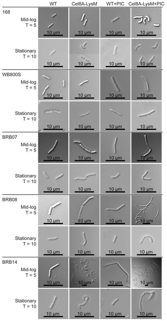
Figure 4:

Differential interference contrast (DIC) microscopy of reporter protein expressing strains. Images were acquired for the parent strain (WT), cells expressing Cel8A-LysM (Cel8A-LysM), the parent strain grown in media containing a protease inhibitor cocktail (WT + PIC), and cells expressing the reporter protein grown in media containing a protease inhibitor cocktail (Cel8A-LysM + PIC). Images of mid-log and stationary phase cells were collected at T = 5 and T = 10 hours, respectively. Scale bar is presented at the bottom of each image.
