Figure 3.

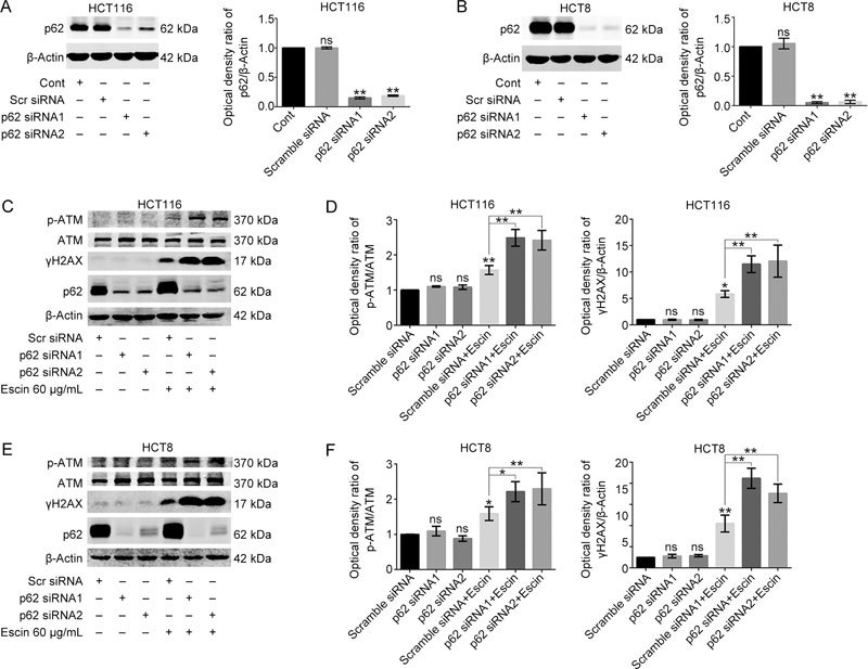
p62 protected DNA from escin-induced DNA damage. (A–B) Knockdown efficiency of p62 in HCT116 and HCT8 cells. Cells were transiently transfected with scramble siRNA and p62 siRNA1/2 for 36 h. The expression level of p62 was tested by Western blotting. Quantitative analysis of the optical density ratio of p62 compared with β-actin is shown. (C–F) Expression of DNA damage related proteins after p62 knockdown with escin treatment. Cells were treated with 60 μg/mL escin for 12 h after scramble siRNA and p62 siRNA1/2 transfection for 36 h. The protein levels of p-ATM, ATM, γH2AX, p62 and β-actin were detected by Western blotting (C and E). Quantitative analysis of the optical density ratio of p-ATM and γH2AX compared with β-actin is shown (D and F).
