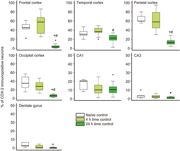
Figure 1.

The ratio of neocortical COX-2-immunopositive neurons decreased over time under normoxic conditions in anesthetized time control animals. In the different cortical lobes, the percentages of COX-2-immunopositive cells determined in samples from naïve animals (brains harvested immediately after anesthesia, n=5) and from normoxic time controls with 4 h of survival after anesthesia (n=12) displayed similar COX-2 expression in accordance with previous data showing regional differences22. However, the percentage of COX-2-immunopositive neurons in the 24 h survival time control group (n=14) was significantly reduced compared with those of both the naïve and the 4 h time control groups in all neocortical regions. Interestingly, this reduction did not affect the CA1 and CA3 hippocampal subfields. (* P<0.05 vs naïve animals, # P<0.05 vs 4 h time control; ANOVA on ranks, Dunn's post hoc test. Bold line, box, and whiskers represent the median, 25th–75th, and 10th–90th percentiles, respectively; black dots are outliers).
