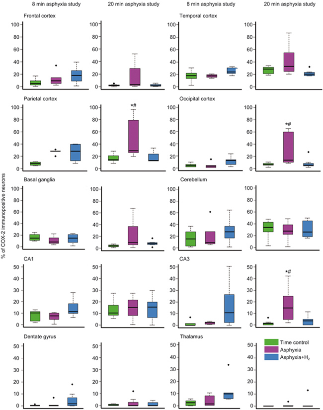
Figure 2.

The effect of perinatal asphyxia and neuroprotective molecular H2 on the ratio of COX-2-immunopositive neurons determined after 24 h of survival. Two levels of perinatal asphyxia of different durations (8 and 20 min) were studied, and both asphyxia groups (n=7-7) were compared with the corresponding time control (n=7-7) and H2-treated asphyxia groups (n=7-7). In the first study using 8-min asphyxia, neuronal COX-2 expression was not significantly affected in either the asphyxia or the H2-treated asphyxia group. On the other hand, in the second study, 20-min asphyxia elicited significant elevations in the ratios of COX-2-immunopositive neurons in the parietal and occipital cortices and in the hippocampal CA3 region. The trends towards increased COX-2 expression were also observed in the frontal and temporal cortices and the basal ganglia, although these differences did not reach statistical significance in these regions. The hippocampal CA1 region, dentate gyrus, and thalamus showed low immunopositivity, which was unaffected by both asphyxia and H2 treatment. Approximately one-third of cerebellar Purkinje-cells were COX-2-immunopositive, and no marked differences were detected among the three groups. (* P<0.05 vs time control. # P<0.05 vs asphyxia+H2; ANOVA on ranks, Student-Newman-Keuls post hoc test. Bold line, box, and whiskers represent the median, 25th–75th, and 10th–90th percentiles, respectively; black dots are outliers).
