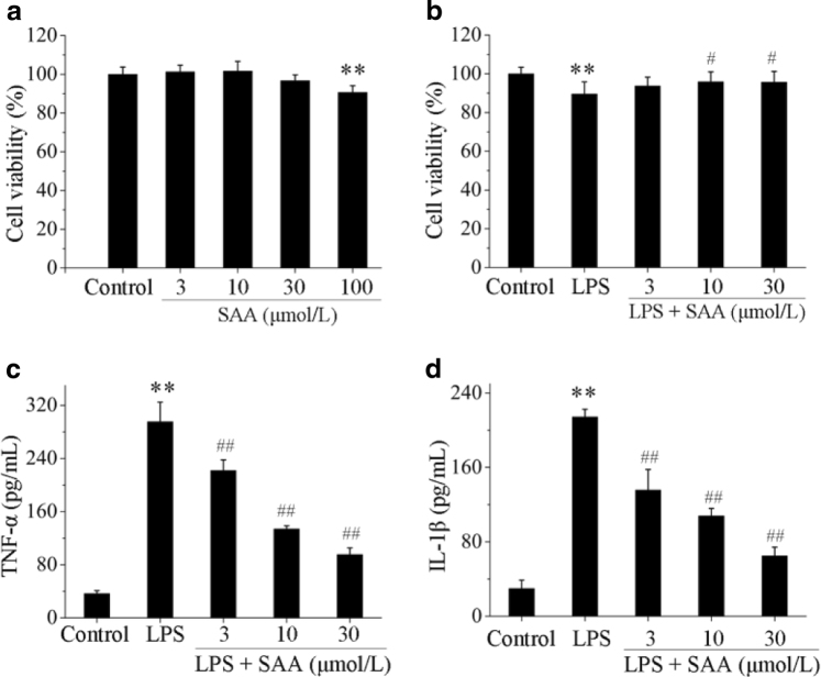
Fig. 3.
The effects of SAA on HK-2 cell viability and TNF-α and IL-1β levels in LPS-induced HK-2 cells. Cells were pretreated with or without various concentrations of SAA for 2 h, and then co-incubated with or without LPS (5 µg/mL) for 24 h. Cell viability was assessed by MTT assay. Effects of SAA on HK-2 cell viability (a) and effects of SAA on LPS-induced HK-2 cell viability (b). The levels of the pro-inflammatory cytokines TNF-α and IL-1β in each group were measured by ELISA. TNF-α (c) and IL-1β (d) levels in LPS-induced HK-2 cells. Data are expressed as the means ± SD (n = 3). **P < 0.01 vs. control group; #P < 0.05, ##P < 0.01 vs. LPS group

