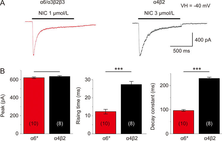
Figure 5.

Kinetics of α6*-nAChR-mediated whole-cell current in human SH-EP1 cells. EC50 (1 μmol/L for α6*-nAChR, (A), or 3 μmol/L for α4β2-nAChR, (B) concentrations of nicotine were applied to induce whole-cell inward currents in transfected SH-EP1 cells stably expressing either the human α6*-nAChR (A) or the human α4β2-nAChR (B). (C) Bar graphs summarizing results of replicate studies (10 cells for α6*-nAChR and 8 cells for α4β2-nAChR) illustrating differences between the α6*-nAChR (red columns) and the α4β2-nAChR (black columns) nAChR responses in the rising times of whole-cell currents (B, middle panel) and whole-cell current decay constants (B, right panel). There was no significant difference in peak nicotine current amplitudes, but there was a significant difference in the rise time (B, left panel). Data were represented as the means, and vertical bars represent SEMs. Asterisks *** represent significance level P<0.001 between α6*- and α4β2-nAChRs.
