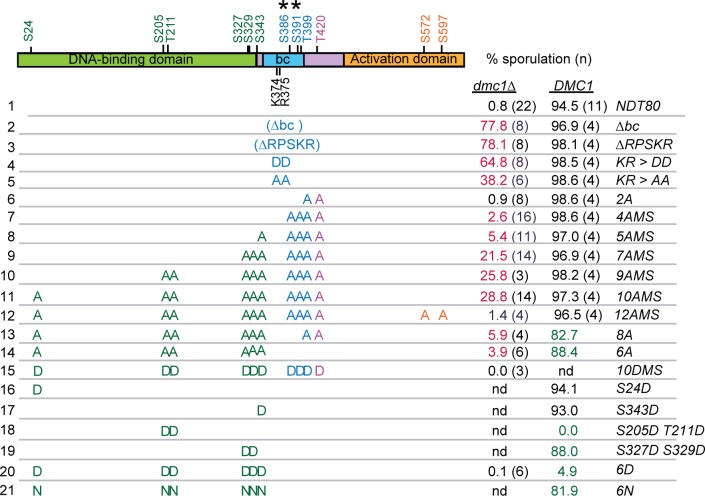
Fig 2. Sporulation analysis of various ndt80 mutants in dmc1Δ and DMC1 diploids.
Potential Mek1 phosphorylation sites are indicated in green, purple and gold, corresponding to the protein domain and have the sequence RXXT/S. Asterisks indicate non-consensus sites detected as phosphorylated in global phosphoproteomic analyses of dmc1Δ- arrested cells [73]. Sporulation was assayed after three days at 30ºC on solid sporulation medium. Each row represents a diploid homozygous for a different allele of NDT80 with mutated residues indicated by either A (alanine), D (aspartic acid) or N (asparagine). “nd” indicates no data. (NDT80, pHL8; NDT80-Δbc, pHL8-Δbc; ndt80-ΔRPSKR, pNH317; ndt80-KR>AA, pHL8-KR>AA; ndt80-KR>DD, pHL8-KR>DD; ndt80-2A, pHL8-2A; ndt80-4AMS, pHL8-4AMS; ndt80-5AMS; pHL8-5AMS; ndt80-7AMS, pHL8-7AMS; ndt80-9AMS, pHL8-9AMS; ndt80-10AMS, pHL8-10AMS; ndt80-8A, pNH405; ndt80-6A, pNH400; ndt80-10DMS, pHL8-10DMS; ndt80-S24D, pHL8-S24D; ndt80-S343D, pHL8-S343D; ndt80-S205D T211D, pHL8-S205D T211D; ndt80-S327D S329D, pHL8-S327D S329D; ndt80-6D, pNH401; ndt80-6N, pHL8-6N. Sporulation was scored in either a dmc1Δ (NH2402) or DMC1 diploid (NH2081). Values in magenta are significantly higher than dmc1Δ NDT80 with p values <9 X10⁻¹⁰, while values in green are significantly lower than DMC1 NDT80 with p values < 0.014 (ndt80-6A) or 9.58 X 10⁻⁸. p values were determined using a one-sided Fisher’s Exact Test. Strains, standard deviations, number of biological replicates and p values are listed in Data S1.

