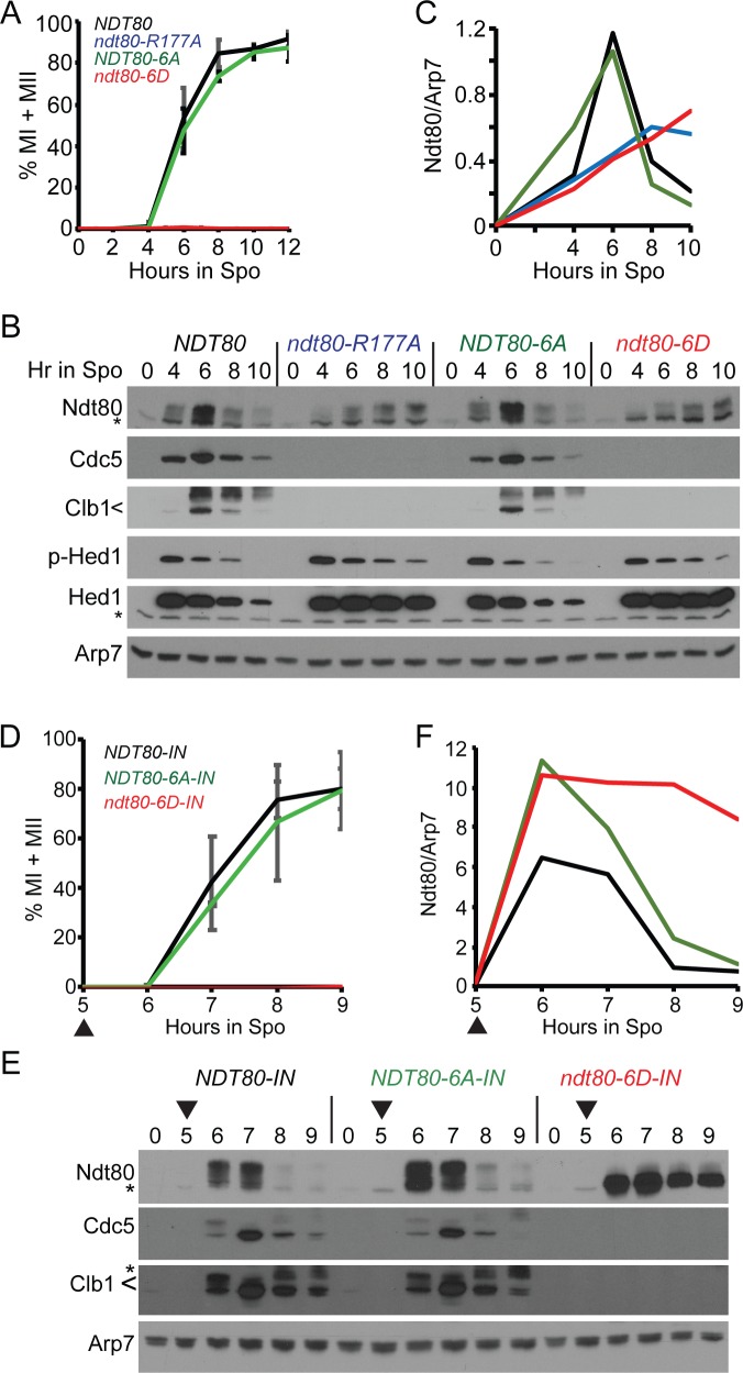
Fig 4. Negatively charged mutations in the Ndt80 DNA binding domain constitutively inactivate Ndt80.
(A-C) Endogenous NDT80. (A) Meiotic progression. Cells expressing NDT80 (NH2426::pEP1052::pHL82), ndt80-R177A (NH2426::pEP1052::pHL8-R177A2), ndt80-6A (NH2426::pEP1052::pNH4002), and ndt80-6D (NH2426::pEP1052::pNH4012) were transferred to Spo medium to induce sporulation. Meiotic progression was determined as described in Fig 3 using three independent timecourses with error bars indicating the standard deviations. (B) Immunoblot analysis of protein extracts from one of the timecourses used in (A). (C) Quantification of Ndt80 signal in (B). Ndt80 was normalized to Arp7 from the same lane on the same gel. The signal from non-specific bands was eliminated by subtracting the Ndt80/Arp7 from 0 hours from each timepoint of the appropriate strain. (D-F) Inducible NDT80. (D) Meiotic progression. Cells expressing NDT80-IN (NH2426::pEP1052::pBG42), ndt80-6A-IN (NH2426::pEP1052::pXC112), and ndt80-6D-IN (NH2426::pEP1052::pXC122) were transferred to Spo medium and incubated for five hours. NDT80 expression was induced by addition of estradiol (ED) to a final concentration of 1 μM (indicated by arrowheads). Data represent the average values of two experiments with error bars indicating the ranges. (E) Immunoblot analysis of proteins extracts obtained from one of the timecourses used in (D). (F) Ndt80 quantification from (E).

