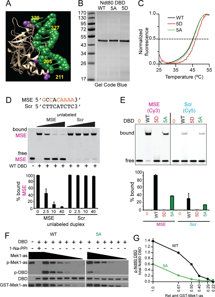
Fig 6. In vitro DNA binding and kinase assays using recombinant Ndt80 DBD proteins.
(A) Structure of Ndt80 DBD bound to a WT MSE [67]. DBD is tan colored and the MSE is green. Putative Mek1 phosphorylated amino acids (numbered in yellow) are indicated in purple. (B) WT, 5A and 5D DBD proteins after purification from E. coli. 800 ng protein from the 100 mM imidazole elution for Ndt80 WT, 5A and 5D DBDs (S1 Fig) were fractionated using a 12% SDS-polyacrylamide gel and stained with Gel Code Blue. (C) Melting temperatures (Tm) were determined by differential scanning fluorimetry (DSF). A representative thermal denaturation curve from one of three independent experiments is shown for each protein. (D) DNA binding assays. Twenty-eight-mer duplexes containing either the nine-base pair MSE from SPS4 or a non-MSE (Scr) sequence were used. MSE consensus sites are indicated in orange. All reactions contained 10 nM Cy3-labeled MSE duplex (magenta) and 50 nM Ndt80 WT DBD. DNA binding specificity was assessed by the addition of unlabeled MSE or Scr duplexes in increasing concentrations (2.5X, 20X or 40X). Reactions were fractionated on native polyacrylamide gels and Cy3 fluorescence detected using a phosphoimager. Quantification was performed on two independent experiments. Error bars indicate the ranges. (E) Comparison of different DBDs binding to MSE or Scr sequences. The indicated Ndt80 DBD (50 nM) was incubated with either 50 nM Cy3-labeled MSE or Cy5-labeled Scr duplex. Quantification was performed on two replicates run on the same gel for each duplex. Error bars indicate the range. (F) In vitro kinase reactions. Reactions contained 40 ng Ndt80 WT or 5A DBD, 100 μM 6-Fu-ATPγS without (-) or with (+) 1 μM 1-Na-PP1. The starting amount of partially purified GST-Mek1-as was 28 ng (= 1) with dilutions of 0.67, 0.50, 0.40, 0.29 and 0.22 as indicated by the black triangles. Thio-phosphorylated proteins were alkylated by the addition of 2.5 mM p-nitrobenzylmesylate and the resulting epitopes detected using thiophosphate ester antibodies (p-GST-Mek1-as and p-Ndt80 DBD). Ndt80 DBDs and GST-Mek1-as were detected by probing the same reactions with α-Ndt80 and α-Mek1 antibodies, respectively. (G) Quantification of the kinase assays shown in Panel F. This experiment was conducted three times with similar results.

