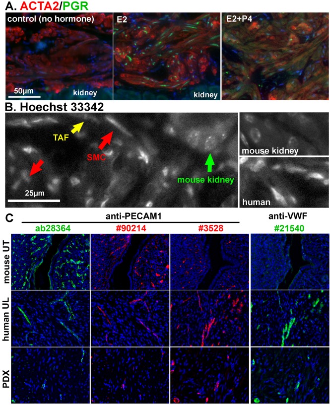
Figure 4. Histological analysis of PDXs.
A. Double immunofluorescence analysis of ACTA2 (red) and PGR (green) in MED12-LM PDXs grown in hosts supplemented with no hormone (control), E2 and E2 + P4. Expression of PGR is E2 dependent. B. Identification of human and mouse cells by nuclear morphology. Human cells have diffuse nuclear staining; mouse cells have granular nuclear staining for heterochromatin. Also indicated are smooth muscle cells (SMC, red arrows) and tumor-associated fibroblasts (TAF, yellow arrows). C. Human origin of vascular endothelial cells in UL PDX. Two anti-PECAM1 (endothelial marker) antibodies, Abcam ab28364 (1:200) and Chemicon #90214 (1:200), preferentially stained mouse uterus (UT) over human ULs, whereas Cell Signaling Technologies #3528 anti-PECAM1 (1:100) antibody preferentially stained human endothelial cells. Chemicon #21540 anti-VWF antibody (1:200) was non-reactive with mouse endothelial cells. Almost all blood vessels in PDXs were strongly positive for VWF, indicating their human origin.

