. 2018 Nov 21;7:e39494. doi: 10.7554/eLife.39494

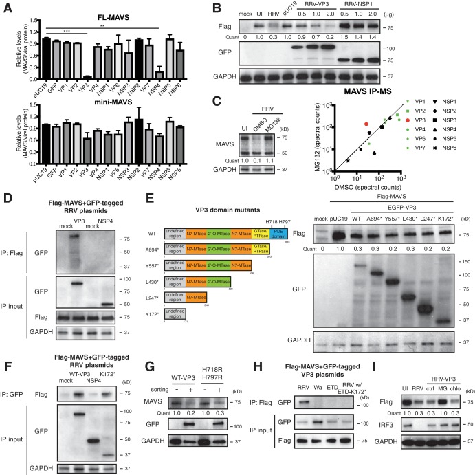
Figure 3. VP3 mediates MAVS interaction and degradation using an N-terminal domain.

(A) MA104 cells were transfected with plasmids expressing each of the 12 GFP-tagged RRV proteins for 72 hr and then subjected to sorting for GFP positive and negative cells. The levels of endogenous full-length and mini-MAVS were normalized to those of GFP-conjugated RRV proteins. (B) MA104 cells were co-transfected with 0.5 μg of Flag-tagged rhesus MAVS and GFP-tagged RRV-VP3 or RRV-NSP1 plasmids for 48 hr. RRV infection (MOI = 1) for 12 hr serves as a positive control. Total cell lysates were harvested and examined by western blot by indicated antibodies. (C) MA104 cells were infected with RRV (MOI = 3) for 8 hr in vehicle control (DMSO) or MG132 (10 μM) treatment. The lysates were subjected to immunoprecipitation using anti-MAVS antibody and analyzed by mass spectrometry for viral proteins. (D) MA104 cells were co-transfected with Flag-tagged MAVS and GFP-tagged RRV-VP3 or RRV-NSP4 for 48 hr and lysates were precipitated with anti-Flag antibody and GFP levels were examined by western blot. (E) Schematic diagram of WT and mutant VP3 proteins with defined domains illustrated in colors and catalytic sites of phosphodiesterase (PDE) activity indicated (left panel). MA104 cells were co-transfected with Flag-tagged MAVS and GFP-tagged VP3 mutants for 48 hr. The lysates were harvested and the levels of Flag and GFP were measured by western blot. (F) MA104 cells were co-transfected with Flag-tagged MAVS and GFP-tagged RV proteins (WT VP3, N-terminal K172* VP3, and NSP4) for 48 hr and lysates were harvested for immunoprecipitation using anti-GFP antibody. (G) MA104 cells were transfected with GFP-tagged wild-type or PDE mutant RRV-VP3 for 72 hr and then subjected to sorting for GFP positive cells. The levels of endogenous MAVS and GFP were examined by western blot. (H) MA104 cells were co-transfected with Flag-tagged MAVS and GFP-tagged VP3 from RRV (simian), Wa (human), ETD (murine) RV strains or chimeric RRV VP3 with 171 amino acids from ETD VP3, treated with MG132. The lysates were harvested for immunoprecipitation using anti-Flag antibody and probed for GFP levels. (I) MA104 cells were co-transfected with Flag-tagged MAVS and GFP-tagged RRV-VP3, and treated with MG132 (MG) or chloroquine (chlo) for 12 hr. The levels of Flag and IRF3 were examined by western blot. For all figures except (c), experiments were repeated at least three times with similar results. Experiments in (c) were performed once. Data are represented as mean ± SEM. Statistical significance is determined by Student’s t test (*p≤0.05; **p≤0.01; ***p≤0.001).

Figure 3—source data 1. Source data for Figure 3C: MAVS-RV protein IP counts.
DOI: 10.7554/eLife.39494.008

Figure 3.

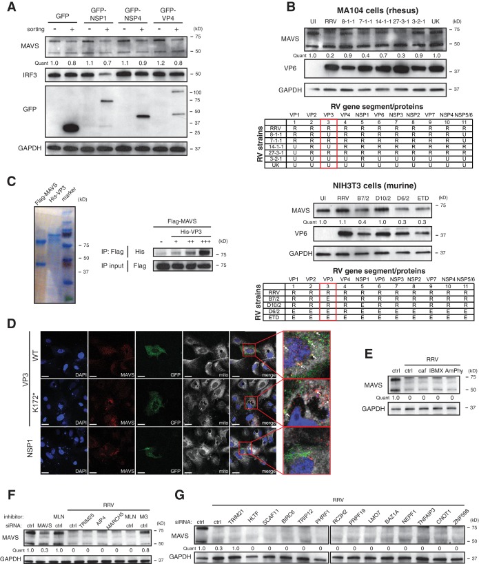
Figure 3—figure supplement 1. VP3 localizes to the mitochondria and induces MAVS degradation.

Figure 3—figure supplement 1.

(A) MA104 cells were transfected with GFP-tagged RRV NSP1, NSP4 or VP4 for 72 hr and subject to sorting for GFP positive and negative cells. The levels of MAVS, IRF3, GFP and GAPDH were determined by western blot. Note that RV VP4 is cleaved into N-terminal VP8* and C-terminal VP5*. GFP tags are constructed on the amino terminus, hence giving the larger band (GFP-VP4) and the smaller band (GFP-VP8*). (B) MA104 cells were infected with reassortant RVs between simian RRV and bovine UK strains (MOI = 3) for 12 hr (top panel). NIH3T3 cells were infected with reassortant RVs between simian RRV and murine ETD strains (MOI = 3) for 12 hr (bottom panel). The levels of MAVS, VP6 and GAPDH were measured for both experiments. The genetic reassortment in different RV strains was illustrated in the table (R: RRV; E: EW). The red box highlights the RV gene three that encodes viral protein VP3. (C) Recombinant Flag-tagged MAVS and His-tagged VP3 proteins were purified from CHO-S cell lines and examined by Coomassie staining. Immunoprecipitation was performed using purified Flag-MAVS and an increase dose of His-VP3 with an anti-Flag antibody. The pull-down lysates and IP input were examined by western blot using anti-Flag and anti-His antibodies. (D) MA104 cells were transfected with GFP-tagged RRV VP3 (WT or N-terminal mutant K172*) or RRV NSP1 for 48 hr. The localization of MAVS and viral proteins were examined by confocal microscopy. Insets are enlarged by red boxes and co-localization of MAVS and viral protein is presented as yellow dots, as indicated by white arrows in the merged panel. Scale bar: 10 μm. (E) MA104 cells were infected with RRV (MOI = 3) for 12 hpi in the presence of phosphodiesterase inhibitors. (caf: caffeic acid; IBMX: 3-isobutyl-1-methylxanthine; AmPhy: aminophylline). (F) MA104 cells were transfected with siRNA against indicated E3 ubiquitin ligases for 48 hr and infected with RRV (MOI = 3) for 12 hr with or without inhibitor treatment (MLN: MLN4924; MG: MG132). The lysates were harvested and examined by western blot for the levels of MAVS and GAPDH. (G) MA104 cells were transfected with siRNA against indicated MAVS-interacting host proteins 48 hr and infected with RRV (MOI = 3) for 12 hr. The levels of MAVS and GAPDH were measured by western blot. For all figures, experiments were repeated at least three times.