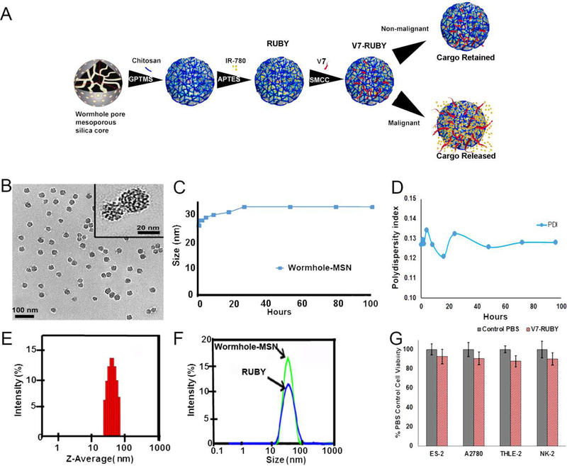
Figure 1. Characterization of the wormhole mesoporous silica nanoparticles.
A) Schematic illustration of the formation of RUBY. B) Transmission electron micrograph (TEM) images with an average particle size of 25 ± 3.1 nm. C) Dynamic light scattering (DLS) of the Wormhole-MSN at multiple timepoints during the synthesis.D) Polydispersity Index (PDI) shows narrow polydispersed distribution as the PDI ranged from 0.12–0.135 over the course of 100 h. E) Dynamic light scattering indicates the average size of the Wormhole-MSN is 33.03 nm at 24 h. F) Chitosan conjugation increased the size of the (RUBY) particle to 38.87 ±1.5 nm. G) In vitro toxicity evaluation of V7-RUBY in ovarian tumor cell lines, non-malignant hepatocytes, and proximal tubule kidney cells was evaluated.Treatment with 0.66 mM empty V7-RUBY particles did not exhibit cytotoxicity on ovarian tumor cells, non-malignant hepatocytes, or proximal tubule kidney cells in comparison to PBS treated controls (p=0.99,0.99,0.98,0.99).

