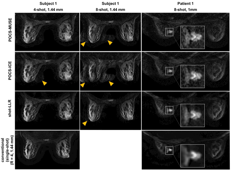
Fig. 5.
Breast images of a volunteer (1st and 2nd columns) and a patient (3rd column) (slice thickness = 4 mm, b-value = 600 s/mm2, nex = 2) reconstructed by different methods under different numbers of shots and different in-plane resolutions. The last row shows the results using the conventional method, in which single-shot and parallel imaging were used, and the reduction factor was 4. Yellow arrows highlight aliasing artifacts. In the 3rd column, an enlarged view of the tumor is provided in the white boxes. Note the improved depiction of the lesion detail in shot-LLR vs conventional single-shot DWI, with reduced artifacts compared to POCS-ICE and POCS-MUSE.

