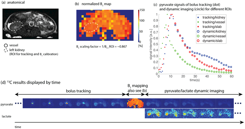
Figure 4:
Results of a hyperpolarized [1-13C]pyruvate study in a normal rat using the proposed method (Fig. 1) with a birdcage coil. Real-time center frequency calibration was not performed in this study. The ROI for both bolus tracking and B1 calibration was on the left kidney. Injection time was 8 s and initial power was purposely set to 120% of the calibrated power in pre-scan. Sequence parameters are presented in Table 1. (a) Proton localizer. (b) Normalized 13C B1 map. Real-time B1 scaling factor (~0.87) matched up with the initial transmit power (120%). (c) Normalized pyruvate signal curves in different ROIs. The ROI (left kidney) bolus peak was successfully detected. (d) 13C results displayed in the order of time. Every other timeframe is shown. The full set of images can be found in Sup. Fig. S1. Experiment recording: https://youtu.be/CN3mIrzmBT8.

