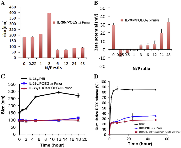
Figure 3.
Particle sizes (A) and zeta potentials (B) of IL-36γ plasmid/POEG-st-Pmor complexes formed at different N/P ratios. Data are expressed as means ± s.e.m. (n=3). (C) The stability of DOX+IL-36γ plasmid/POEG-st-Pmor was examined by incubating complexes (1 mg DOX/mL in PBS, pH 7.4) with bovine serum albumin (BSA, 30mg/mL) at 37°C. Changes in sizes of the complexes over incubation time were followed by DLS. (D) In vitro drug release profiles of DOX from free DOX, DOX/POEG-st-Pmor, and DOX+IL-36γ plasmid/POEG-st-Pmor in PBS at 37°C. Data are mean ± s.e.m. (n=3).

