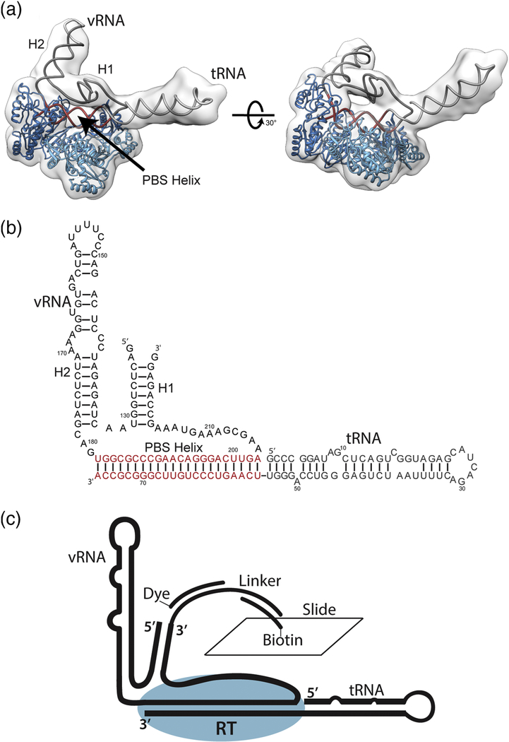
Figure 2.
Global architecture of the +1 HIV-1 reverse transcriptase initiation complex. (a) The vRNA and tRNA form an extended PBS helix which occupies the RT binding cleft. H1 and the elongated tRNA protrude from the polymerization active site and RNase H domain respectively. H1 rests on top of the occupied RT binding cleft. (b) Secondary structure of the vRNA and tRNA as fit to cryo-EM electron density maps. (c) Schematic of constructs used for surface immobilization for smFRET experiments.

