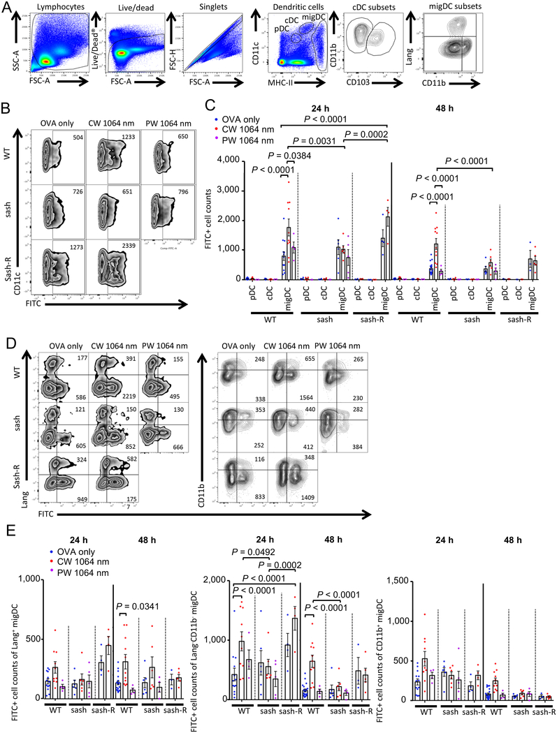
Figure 2. Effect of the NIR laser on DC migration in skin.
The effect of the NIR laser on migration responses of migratory DC (migDC) subsets in skin. Mice were painted with 0.5 % FITC solution on the flank skin 4 hours before vaccination with 40 μg of OVA with or without the NIR laser treatment. Skin-draining lymph nodes (dLN) were analyzed by flow cytometry. A, Gating schematic to identify DC subsets within skin-dLN. Cell counts of B–C, DC subsets, D–E, migDC subsets in the skin-dLN 24 and 48 hours after OVA vaccination are shown. A–D, n= 13, 10, 4, 6, 6, 5, 4, 4 at 24 hours and n= 20, 11, 4, 6, 6, 4, 3, 5 at 48 hours for OVA only in WT, CW 1064 nm in WT, PW 1064 nm in WT, OVA only in sash, CW 1064 nm in sash, PW 1064 nm in sash, OVA only in sash-R, CW 1064 nm in sash-R groups, respectively. Results were pooled from 6 independent experiments and analyzed using two-way ANOVA followed by Tukey’s HSD tests.

