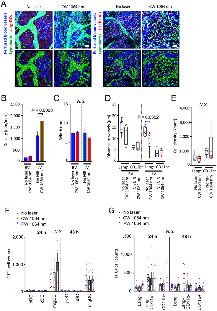
Figure 7. The effect of the NIR laser on microcirculation and DC migration without antigen challenge in skin.
A–E, Morphometry of the vascular and lymphatic networks in skin with or without the NIR laser treatment. Morphological changes of the vascular and lymphatic networks in skin were measured in stacks of the 3D confocal images using Imaris software. The depilated mouse ears were treated with the NIR laser, and the whole-mount ear tissue was stained and imaged for blood vessels, lymphatics, and DC subsets 6 hours after the treatment. A, Representative confocal images of mouse ear stained for blood vessels, lymphatics and DCs 6 hours following the CW NIR laser treatment. Bar = 40 μm. B–C, Morphometry of blood and lymphatic vessels and DCs in skin using Imaris software. Two-way ANOVA followed by the Tukey’s HSD tests. a–c, n= 17, 17 for nor laser control and CW NIR laser-treated groups, respectively. D, Distances between vasculature and DCs, E, Density of Lang+ and CD11b+ DCs in skin. n= 5–8, 5–8 for no laser control and CW NIR laser-treated groups, respectively. Two-way ANOVA followed by the Tukey’s HSD tests. A–E, Results were pooled from 5 independent experiments. F–G, The effect of the NIR laser on the migration response of migDC subsets without antigen challenge in skin. Mice were painted with a 0.5 % FITC solution on the flank skin 4 hours before the NIR laser treatment without any antigen challenge. Cell counts of F, DC subsets G, migDC subsets in the skin-dLN 24 and 48 hours after FITC painting are shown. F–G, n= 13–14, 7, 7 for no laser, CW 1064 nm, PW 1064 nm groups, respectively. Two-way ANOVA followed by Tukey’s HSD tests. Results were pooled from 4 independent experiments.

