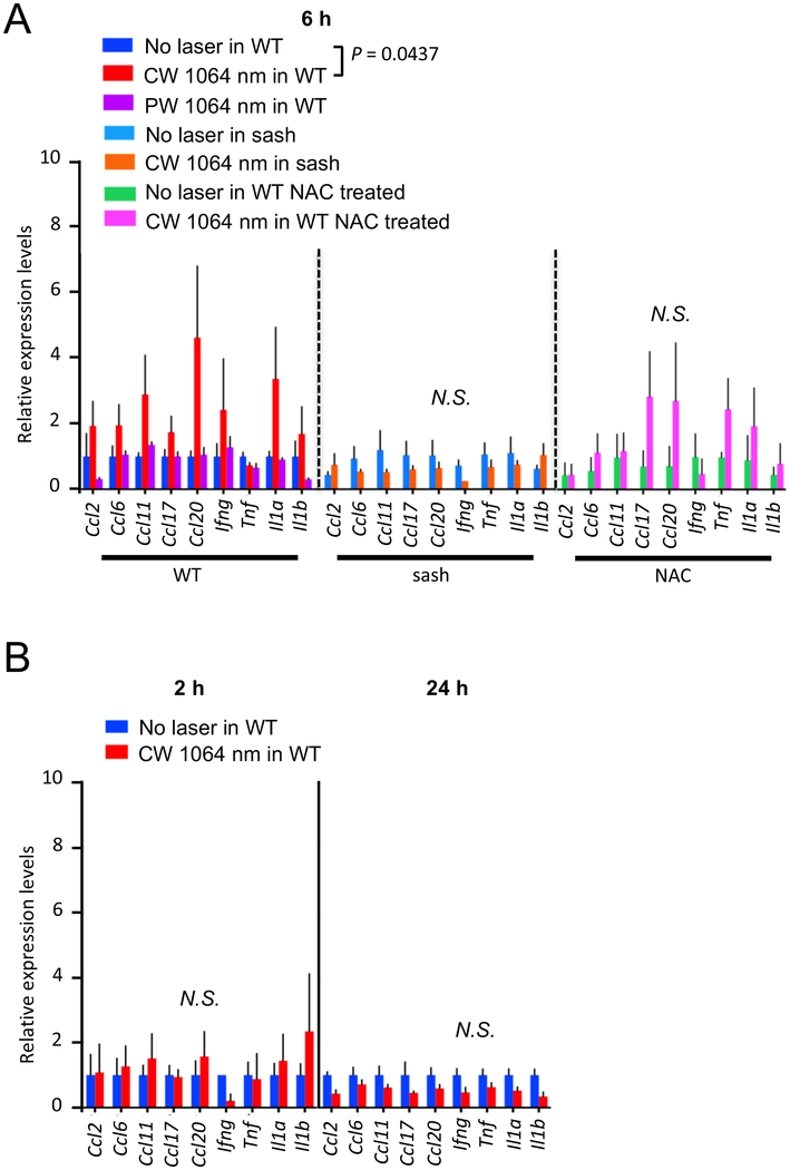
Figure 8. A critical role of MCs and ROS in the chemokine expression in skin in response to the NIR laser.
The effect of the NIR laser on the chemokine expression in skin was evaluated in WT and sash mice. The role of ROS in the chemokine expression in skin in response to the CW NIR laser was also evaluated in NAC-treated WT mice. For antioxidant treatment, mice were treated with NAC for 4 consecutive days before the NIR laser treatment as described above. A, The expression of chemokines in the mouse back skin was measured 6 hours following the CW NIR laser treatment in (left) WT, (middle) sash, and (right) NAC-treated WT mice using qPCR. n = 7, 7, 4, 3, 3, 3, 3 for no laser control in WT, CW and PW 1064 nm laser-treated in WT, no laser control in sash, CW 1064 nm laser-treated in sash, NAC-treated only, CW 1064 nm in NAC-treated groups, respectively. B, The expression of chemokines in the mouse back skin in (left) 2 and (right) 24 hours after the NIR laser treatment in WT mice. n = 4, 4 at 2 hours and n = at 24 hours for no NIR, CW 1064 nm groups. A–B, Student’s t test with stepdown bootstrap and false discovery rate corrections followed by a multivariate discriminant analysis of the 9 genes was used as appropriate. Error bars show means ± s.e.m.

