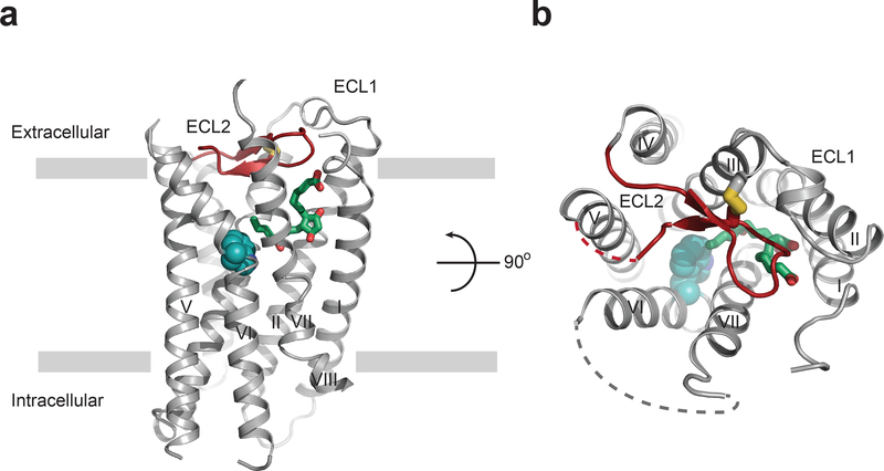
Figure 1|. Overall structure of EP3 receptor bound to misoprostol-FA.
(a) EP3 receptor viewed parallel to the plasma membrane, and (b) from the extracellular side looking down into the misoprostol-FA binding pocket. Misoprostol-FA (green carbon) is shown as sticks. Disordered parts of loops are represented as dashed lines. ECL2 is shown in red. The canonical ‘toggle-switch’ Trp2956.48 is shown with cyan atom spheres for reference.

