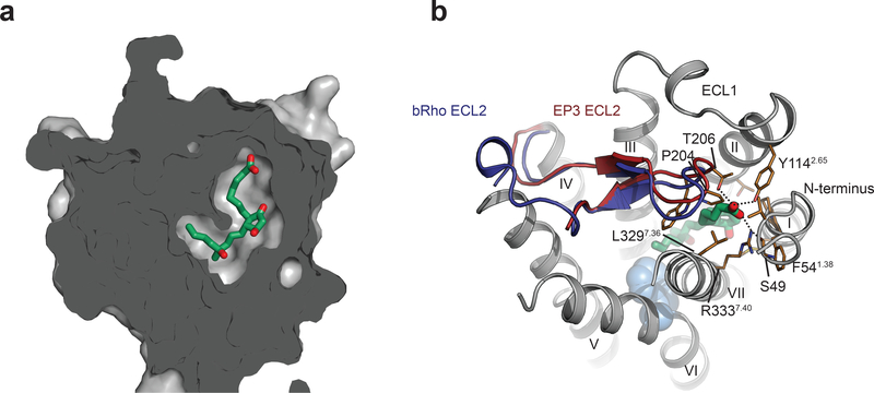
Figure 3|. The binding pocket of misoprostol-FA in EP3 receptor is totally enclosed.
(a) Sliced surface representation of the misoprostol binding pocket. (b) Extracellular view of EP3 receptor with its ECL2 (red) overlaid with the one of bovine rhodopsin (dark blue) (PDB code 1gzm). Trp2956.48 is shown with cyan spheres for reference. Misoprostol-FA is shown as green sticks. EP3 residues are shown as gold stick. Polar interactions are shown as dotted lines.

