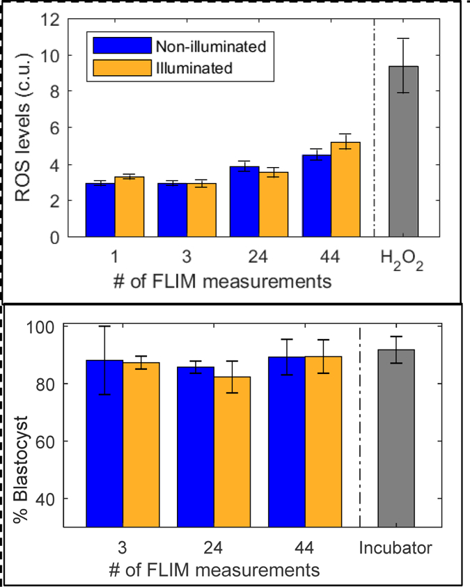
Figure 4:

Evaluation of safety for varying photodoses of FLIM illumination. Top: Reactive oxygen species levels were measured via HC-DCFDA fluorescence (custom units). Significant differences between illuminated and non-illuminated embryos were not observed for any of the photodoses studied. Embryos exposed to 30mM H2O2 were measured as a positive control. Error standard error bars represent variation between individual embryo measurements. Bottom: Embryos were cultured on the microscope, and blastocyst development rates of illuminated embryos were compared to non-illuminated embryos in the same dish. Embryos cultured in a standard incubator were used as a control. FLIM illumination did not have any significant impact on blastocyst development rates. Standard error bars represent variation between experiment batches.
