
Figure 4. period-AID-EGFP flies functional normally on the molecular level.
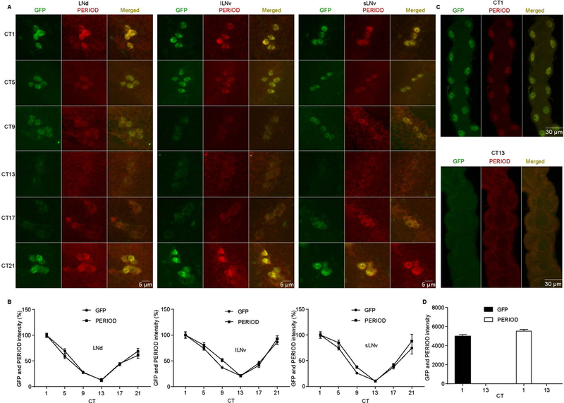
(A) Oscillation of GFP and PER in period-AID-EGFP flies. Adult brains were dissected at CT 1, 5, 9, 13, 17, and 21 at DD1 and circadian neurons were immunostained with anti-GFP and anti-PER. (B) Quantification of GFP and PER average intensity in each clock cell group (n=10). Signal was normalized to the value at CT1 which was set as 100%. CT is indicated on the x axes. (C) Malpighian tubules from period-AID-EGFP flies collected at the indicated circadian time on DD1 were immunostained with anti-GFP and anti-PER. (D) Quantification of GFP and PER intensity in Malpighian tubules (n=8). CT is indicated on the x axes. LNd, dorsal lateral neuron; lLNv, large ventral LN; sLNv, small ventral LN.