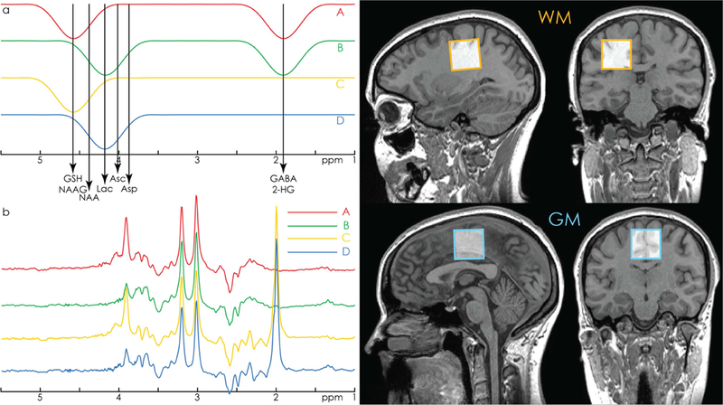
Figure 1.

a. HERCULES editing scheme with editing lobes at 1.9 ppm (targeting GABA and 2- HG), 4.18 ppm (targeting Asp, Asc, Lac), and 4.58 ppm (targeting GSH and NAAG). The 4.58 and 4.18 ppm editing lobes are arranged symmetrically around the 4.38 ppm resonance of NAA. This design minimizes the contribution of highly concentrated NAA to the A-B+C-D difference spectrum, improving the segregation of the aspartyl signals of NAA and NAAG into orthogonal difference spectra. b. Example in vivo HERCULES sub-spectra A, B, C, and D, as produced by the editing scheme in 1a. Residual water signal has been filtered out. c. Example voxel placement in white-matter- and grey-matter-rich regions.
