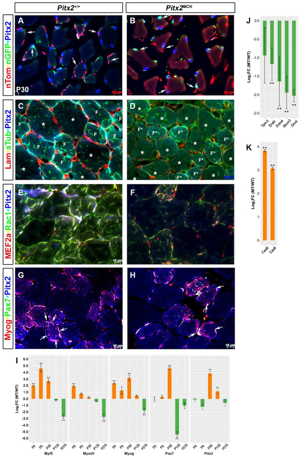
Figure 3. Cytoskeletal Defects Localized to Smaller Fibers in Pitx2MCK muscles.
Immunohistochemical and qPCR comparison of P30 TA muscle Triple labelling to monitor (A, B) docking, conditional allele excision, and Pitx2 expression in myofibers; (C, D) Laminin, alpha-tubulin, and Pitx2 proteins; (E, F) MEF2a, Rac1 and Pitx2 proteins; (G, H) Myog, Pax7, and Pitx2 proteins. Note that smaller (F, F*) and larger(*) fibers respond differently to mutation (see C, D). (I) qPCR analysis of total RNA from TA muscles at P0, P5, P30, P120, and P270. using primers for RNAs encoding muscle specific SSTFs. (J, K) qPCR analysis of P30 TA muscle RNA for cytoskeletal components Tpm3 (Tropomyosin), Enah (Enabled homolog), Dctn4 (Dynactin), Stmn3 (Stathmin), Dmd (Dystrophin), and apoptosis proteins (Caspases 8 and 9).

