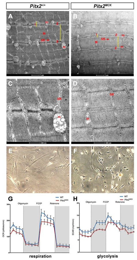
Figure 4. Mitochondrial Loss and Respiration Defects in Pitx2MCK Muscle.
(A, D) Transmission electron microscopy for TA at P30 indicated distorted and smaller sarcomeres. Z, I, and M bands show differences. Mitochondria (M) and sarcoplasmic reticulum (SR) are vestigial. (E-H) Seahorse assay on primary TA muscle cell cultures (E, F) from WT and Pitx2MCK mice (n=3). (G) OCR and (H) ECAR measure mitochondrial respiration and glycolysis, respectively.

