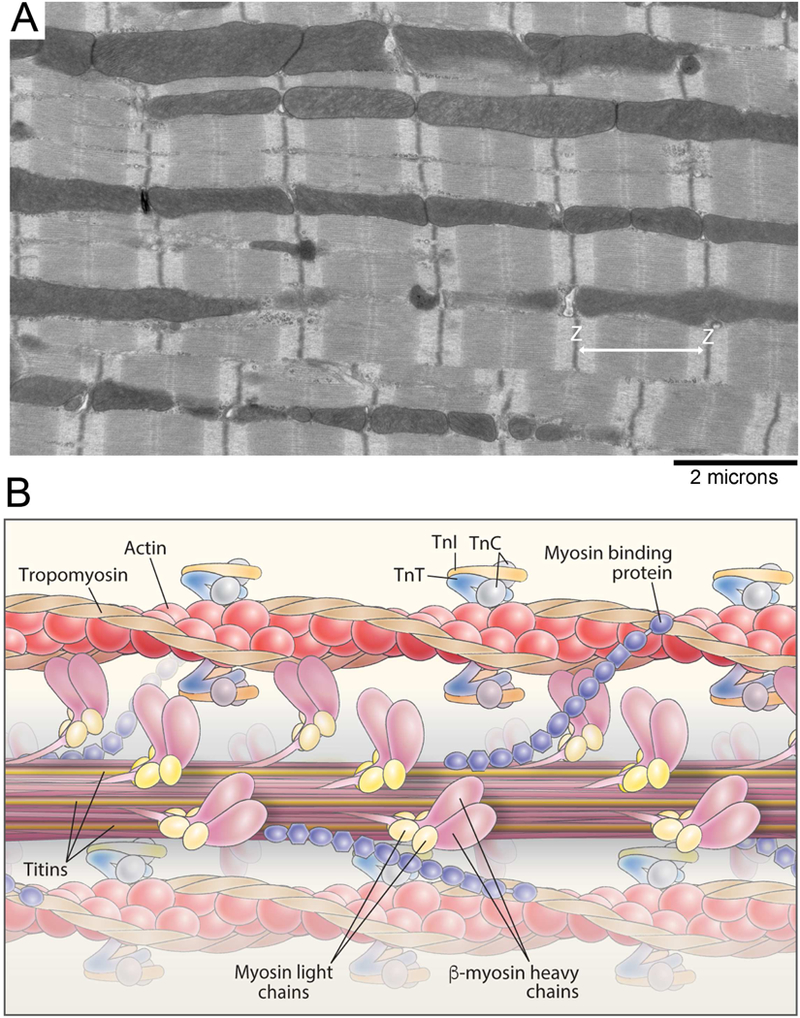
Figure 2. Striated muscle ultrastructure and the myofilament.

A, Electron micrograph of the cardiac myofilament. B, Schematic diagram illustrating the structure of the cardiac myofilament. Myofilament proteins, such as thick filament proteins (titin, myosin, myosin binding protein-C, light chains) and thin filament proteins (actin, tropomyosin, troponin I, C and T), are shown.
