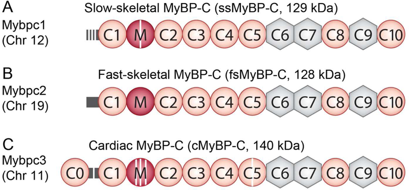
Figure 3. Domain structure of the three paralogs of MyBP-C.

Immunoglobulin domains are shown in orange circles, while fibronectin-III domains are shown in grey hexagons. The proline-alanine (P/A)-rich linker is shown in yellow, and the M-domain is shown as a blue circle. Phosphorylation sites are shown as purple stripes. ssMyBP-C is shown in A with its three phosphorylation sites in the P/A and one within the M-domain. B shows the domain structure of fsMyBP-C. cMyBP-C has an additional N-terminal immunoglobulin domain (C0), four phosphorylation sites in the M-domain, one phosphorylation site in the P/A linker, and a 28 novel amino acid insertion in the C5 domain (red stripe), C.
