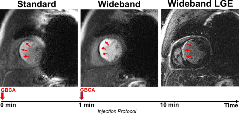
Figure 5.

Example perfusion image sets in a short-axis view in which a perfusion defect is clearly visible. Standard perfusion scan was performed first (left) and shows considerable image artifacts. Wideband perfusion scan was performed second (middle) and shows minimal artifacts. The perfusion defect agrees with myocardial scar shown in wideband LGE (right). A timing diagram displays the injection protocol. GBCA: gadolinium-based contrast agent.
