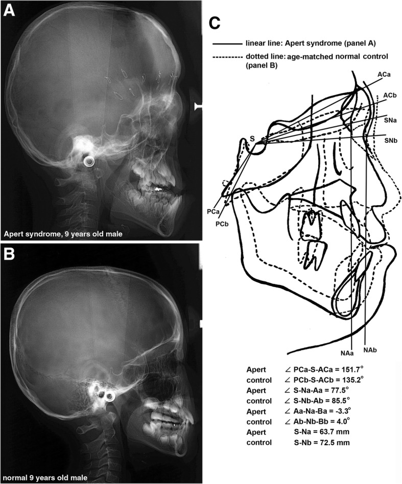
Fig. 4.

Cephalogram: a An Apert syndrome patient, a 9-year-old male, noted with retained suture wires for distraction osteotomy performed at 6 months after birth. b A 9-year-old normal male. c overlapping cephalogram panels a and b by adjusting to the SN line. The Apert syndrome patient showed an increased posterior-anterior cranial base angle (angle PC-S-AC), a retruded maxilla, and a counter-clockwise growth of the mandible compared to the control
