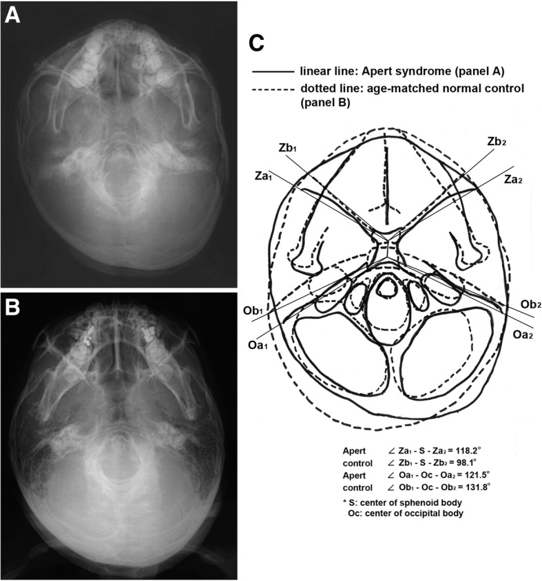
Fig. 6.

Basal cranial view. a An Apert syndrome patient, a 9-year-old male. b A 9-year-old normal male. c Overlapping panels a and b. The Apert syndrome patient showed increased zygomatic axis angle of the cranial base (angle Z1-S-Z2) and decreased otic axis angle of the cranial base (angle O1-Oc-O2) compared to the control
