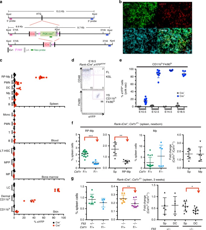
Fig. 4.
Tissue-specific depletion of CSF1R in RP-Mps and their progenitors results in loss of DCs. a Scheme of the generation of Rank-iCre mice (iCre = codon-improved Cre). The iCre-recombinase was knocked into exon 1 of the Rank gene. b Immunohistology on spleen sections from 3-week-old Rank-iCre+;Td-rfp+ mice, ×40 objective was used for picture acquisition, scale bar corresponds to 20 μm. c Lineage tracing of RANK-expressing cells in adult Rank-iCre+;eYFPki/wt mice. LT-HSC long-term HSCs, MPP multipotent progenitors, MP myeloid progenitors, LC Langerhans cells. SD is shown. Gatings are shown in Supplementary Fig. 1a and Supplementary Fig. 3b-d. d Dot plots show the labeling of fetal liver Kit+ Sca-1+ Lineage− (KSL) hematopoietic stem and progenitor cells (top) and yolk sac CD11b+ F4/80hi macrophages (bottom) from E18.5 Rank-iCre+;eYFPki/wt embryos. e Lineage tracing in Rank-iCre;eYFPki/wt embryos gating on CD11blo F4/80hi embryonic macrophages in the yolk sac at the indicated time points. SD is shown. f Plots show the frequencies (left, second right) and fold changes (second left, right) of CD11blo F4/80hi RP-Mps or CD11b+ F4/80lo macrophages and spleen leukocytes from newborn Rank-iCre+;Csf1rF/− and control mice. Two-sided t test (left and second right) and Mann–Whitney U test (right and second right) were performed. SD is shown. g Plots show frequencies and fold changes of DCs and spleen leukocytes from Rank-iCre+;Flt3−/−;Csf1rF/− and control mice at 3 weeks of age. Two-sided t test (left) and Mann–Whitney U test (right) were performed. SD is shown

