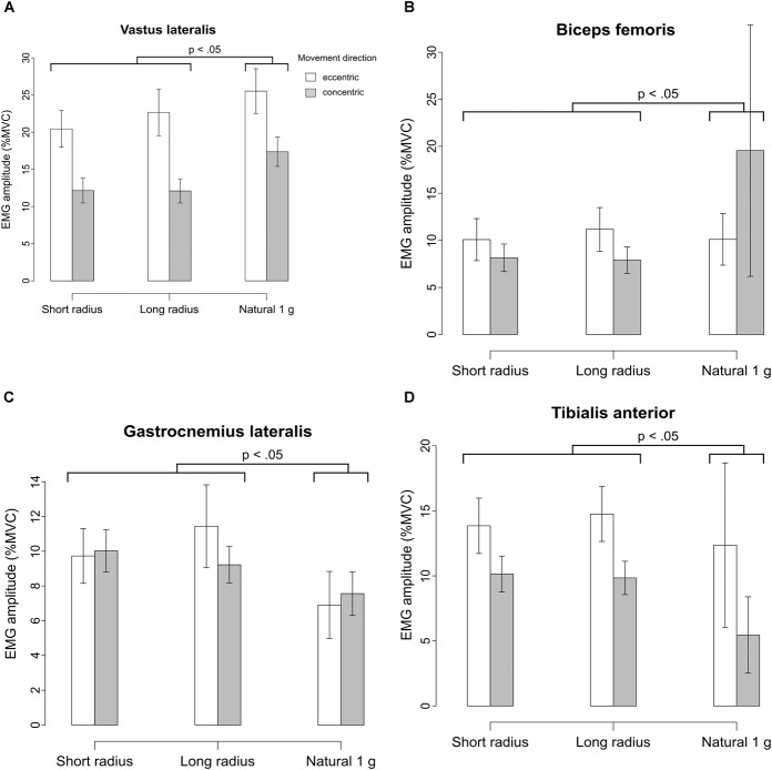
FIGURE 9.
(A) Normalized EMG signals of the left vastus lateralis as a percentage of MVC amplitude during eccentric/concentric contraction under the three g-conditions. Showing mean ± SE. This muscle showed particularly high activation during eccentric movement under all conditions. (B) Normalized EMG signals of the left biceps femoris as a percentage of MVC amplitude during eccentric/concentric contraction under the three g-conditions. Showing mean ± SE. Highest activation was measured during concentric movement under natural gravity. (C) Normalized EMG signals of the left gastrocnemius lateralis as a percentage of MVC amplitude during eccentric/concentric contraction under the three g-conditions. Showing mean ± SE. The muscles of the lower leg were activated more strongly on the centrifuge. (D) Normalized EMG signals of the left tibialis anterior as a percentage of MVC amplitude during eccentric/concentric contraction under the three g-conditions. Showing mean ± SE.

