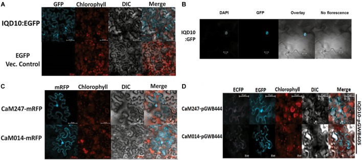
FIGURE 4.
Subcellular localization of PdIQD10, CaM247 and CaM014. (A) Subcellular localization of PdIQD10-GFP was detected in the cytoplasm and plasma membrane. The fluorescence was detected uniformly throughout the cell post 72 h of agroinfiltration of tobacco leaves with the Agrobacterium clone containing PdIQD10 in pGWB405 vector. (B) Repeated assays detected PdIQD10-GFP localization in the nucleus of tobacco leaves agroinfiltrated with the clone harboring the same construct PdIQD10 in pGWB405. (C) CaM247-mRFP and CaM014-mRFP were found to be localized throughout the tobacco cells and in nucleus upon agroinfiltration of clones harboring CaMs in pGWB454 vector. (D) Co-localization of PdIQD1-EGFP with CaM247-CFP or CaM014-CFP was performed by mixing the Agrobacterium cultures in equal (1:1) ratio before agroinfiltration into tobacco leaves. To increase accessibility of these subcellular localization images, the yellow color channel was converted to magenta uniformly across all images in the CMYK color spectrum. The original RGB color scheme images are provided in Supplemental Figure 10. The color scheme is as follows: GFP/RFP: blue/cyan; chlorophyll, FM64 and mCherrry: red/orange; DAPI/CFP: gray/white.

