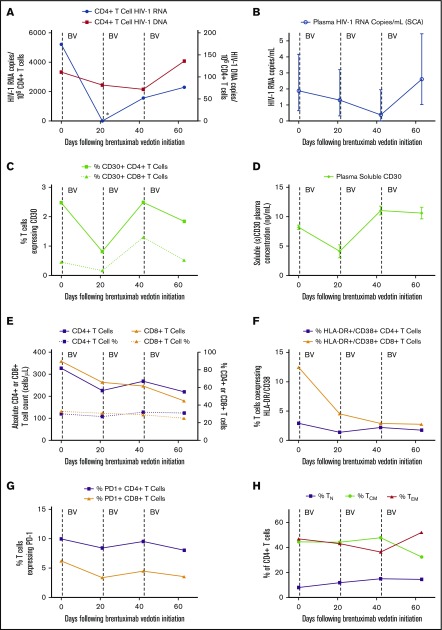
Figure 1.
Changes in measures of HIV persistence, CD30 expression, and immune phenotype during brentuximab vedotin therapy. (A) CD4+ T-cell–associated HIV-1 RNA became undetectable 3 weeks following the first brentuximab vedotin (BV) administration as indicated by the asterisk (*). (B) Residual low-level plasma viremia transiently decreased following anti-CD30 treatment; mean values with 95% confidence intervals from replicate testing in the single-copy assay (SCA) are shown. CD30+CD4+ T-cell percentages (C) and soluble CD30 protein levels in plasma (D) decreased following the initial brentuximab vedotin dose, but increased following the second infusion. Mean and standard deviation are shown for enzyme-linked immunosorbent assay replicate testing. The initial decrease and subsequent rise in HIV-1 RNA and DNA levels tracked with CD30 expression. (E) Absolute CD4+ and CD8+ T-cell counts declined following brentuximab vedotin administration but percentages remained stable throughout treatment. Markers of CD8+ T-cell activation declined following anti-CD30 therapy (F), but minimal or no changes were observed over time in percentages of lymphocytes expressing PD-1 (G). The percentage of CD4+ T cells of naive (TN), central memory (TCM), and effector memory (TEM) phenotype are shown in panel H. Effector memory RA+CD4+ T-cell percentages were all ≤1% and are not shown. Dotted lines represent brentuximab vedotin infusions.

